Cardiac Masses: Tumors and Mimics
Onno Mets¹, Anje Spijkerboer¹, Robin Smithuis²
¹Amsterdam University Medical Center, ²Alrijne Hospital, Leiden, the Netherlands
Publicationdate
Cardiac masses are uncommmon and include a wide range of lesions, such as non-neoplastic entities, primary benign or malignant tumors, and metastases.
Often discovered incidentally, they typically require further imaging for accurate characterization.
Because cardiac lesions are encountered infrequently in clinical practice, establishing a comprehensive differential diagnosis is challenging.
In this article we will discuss:
- Overview of cardiac masses
- Differential diagnostic considerations
- Complementary roles and diagnostic value of the various imaging modalities.
Introduction
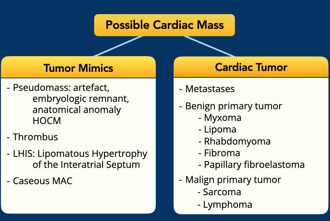
Differential diagnosis
Cardiac structural abnormalities are frequently identified incidentally during echocardiography or CT imaging, irrespective of whether the study was performed to assess cardiac symptoms.
Pseudomass
- The initial diagnostic consideration is to determine whether the finding represent a true cardiac mass, or if it is a pseudomass?
Tumor mimic
- If a true cardiac mass is confirmed, the differential diagnosis is heavily influenced by the patients age and medical history.
- Thrombus or vegetation should be prioritized in the differential, because they are the most common cardiac masses and tumor mimics, especially in the context of myocardial infarctions and indwelling devices (thrombus) or heart valves (vegetation).
Thrombi are most commonly observed in characteristic locations, such as the left atrial appendage or a dilated left ventricle.
Cardiac tumor
- Primary cardiac tumors are exceedingly rare, with the majority of cardiac neoplasms representing metastatic disease.
- The incidence of benign cardiac tumors varies by age:
- Myxoma is the most prevalent in adults (~50%)
- Rhabdomyoma predominates in children (~45%)
Multimodality imaging
Multimodality imaging enhances diagnostic accuracy.
- Ultrasound (TTE/TEE) is first-line: accessible, non-invasive, high spatial/temporal resolution, but operator-dependent and limited by acoustic windows.
- CT offers rapid, high-resolution imaging with excellent assessment of calcification, fat, extent, origin, and vasculature, but involves radiation and contrast.
- MRI provides detailed tissue characterization and functional data, but is limited by availability, scan time, and contraindications (e.g., devices, claustrophobia).
- FDG-PET aids in differentiating malignant/benign lesions and staging, but is costly, involves radiation, and has lower resolution with potential false positives.
- Tissue sampling (biopsy/resection) remains the gold standard for definitive diagnosis when feasible.
Video Example
Multimodality imaging of a malignant cardiac tumor, which proved to be an undifferentiated sarcoma
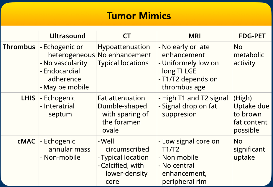
Tumor mimics
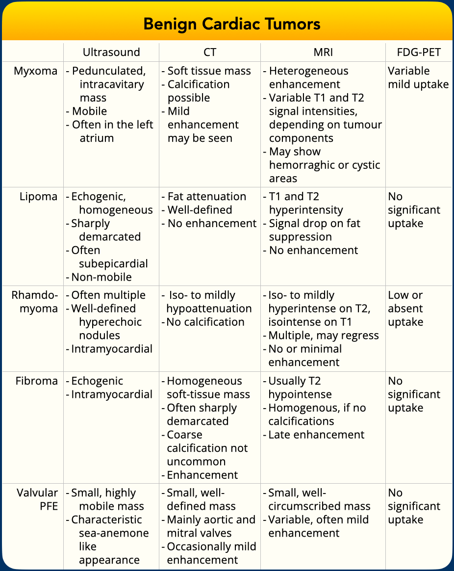
Benign Cardiac Tumors
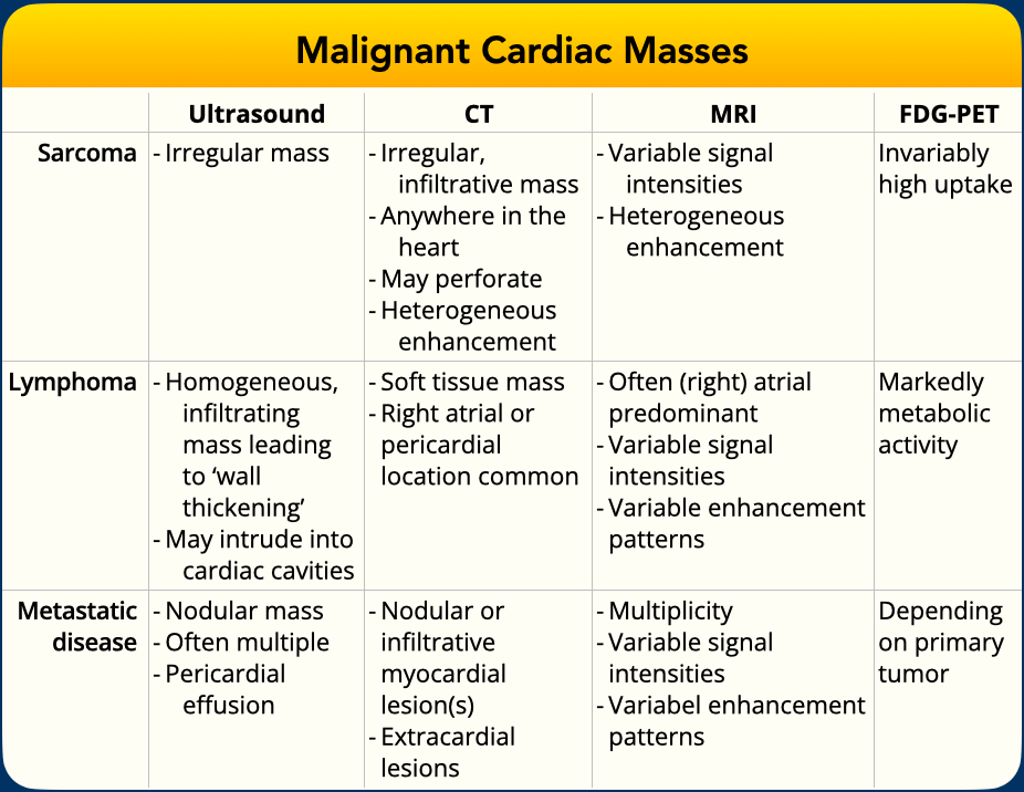
Malign Cardiac Tumors
Tumor Mimics
Pseudomasses
Cor triatriatum sinistrum
Case Presentation
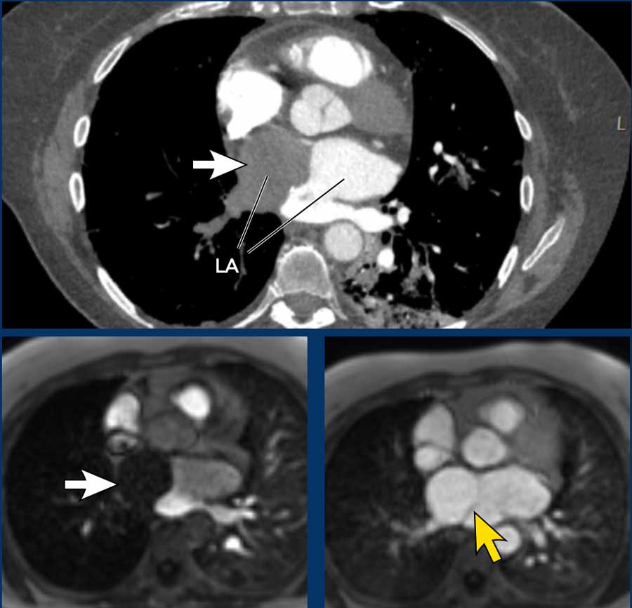
Initial suspicion on CT pulmonary angiogram was for an atrial thrombus or possible cardiac mass (white arrows).
Further evaluation demonstrated delayed filling of the right-sided chamber within a septated left atrium (yellow arrow).
This is consistent with the diagnosis cor triatriatum sinistra.
Cor triatriatum sinistrum is a rare congenital cardiac anomaly characterized by the presence of a fibromuscular membrane that divides the left atrium into two chambers:
- Proximal chamber: Receives pulmonary venous return.
- Distal chamber: Contains the mitral valve and left atrial appendage.
The membrane typically has one or more openings, allowing blood flow between the chambers.
 Obstructive hypertrophic cardiomyopathy (HOCM) with asymmetric myocardial thickening
Pseudomass (2) - Obstructive Hypertrophic Cardiomyopathy
Case presentation
Initial suspicion on CT pulmonary angiogram was for an atrial thrombus or possible cardiac mass (white arrows).
Further evaluation demonstrated delayed filling of the right-sided chamber within a septated left atrium (yellow arrow).
This is consistent with the diagnosis cor triatriatum sinistra.
Cor triatriatum sinistrum is a rare congenital cardiac anomaly characterized by the presence of a fibromuscular membrane that divides the left atrium into two chambers:
- Proximal chamber: Receives pulmonary venous return.
- Distal chamber: Contains the mitral valve and left atrial appendage.
The membrane typically has one or more openings, allowing blood flow between the chambers.
Pseudomass (3) - Thrombus mimic
Case presentation
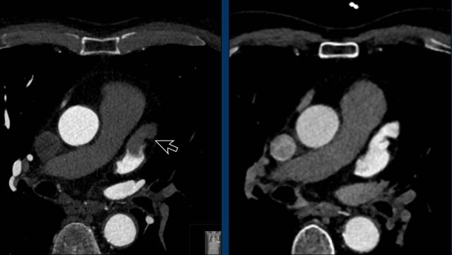
Suspected left atrial
appendage thrombus, which proved not present on a slightly later contrast phase
series.
If present at the scanner, consider a late phase in prone position instead of a separate repeat examination later.
Thrombus
Cardiac thrombi are the most common intracardiac masses, typically forming in the left atrial appendage or left ventricular apex due to blood stasis, endothelial injury, or hypercoagulability. Predisposing conditions include atrial fibrillation, prior myocardial infarction with left ventricular dysfunction, and indwelling cardiac devices/catheters. These thrombi may lead to systemic or pulmonary emboli.
Example
Post-MI, a left apical thrombus may be visualized on 4-chamber cine (yellow arrow). After anticoagulation, CT can show thrombus resolution, with fibrofatty infiltration and myocardial thinning indicating prior infarction (arrowheads).
Images
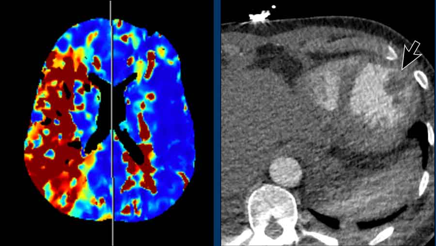
A systemic embolic event due to a left ventricular apical thrombus (arrow) in a patient presenting with acute right M1 occlusion.
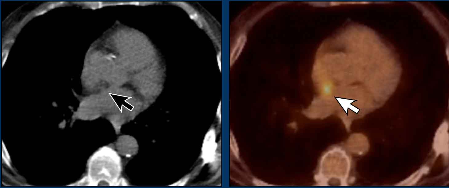
Lipomatous hypertrophy of the interatrial septum (LHIS)
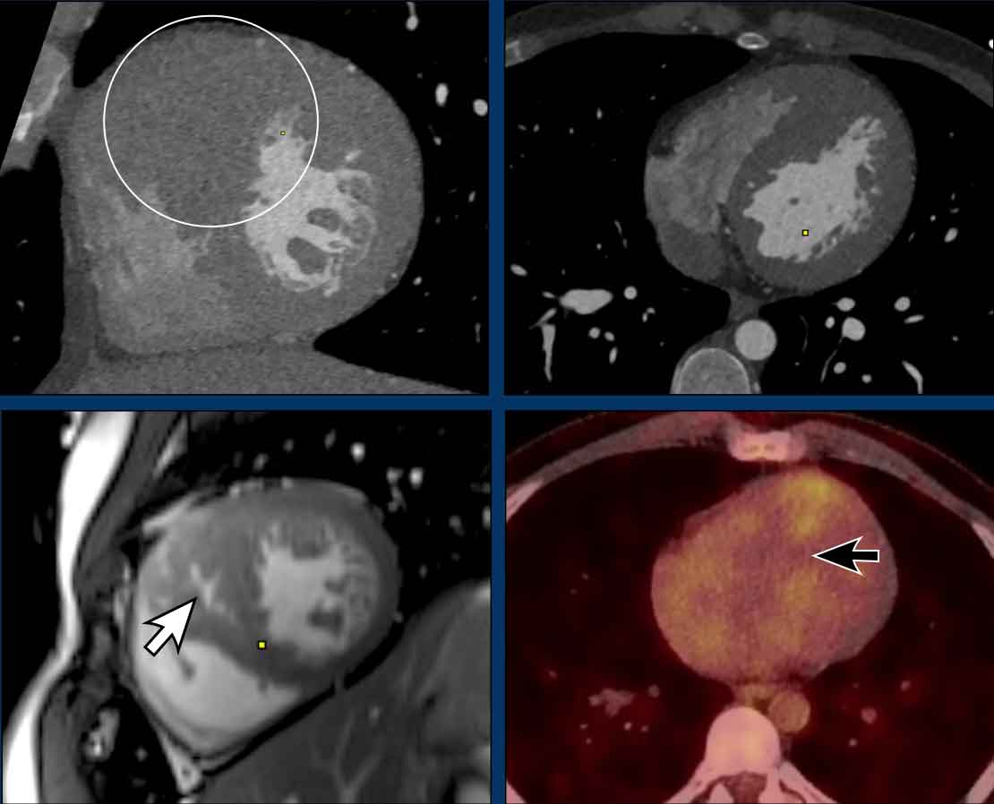
LHIS is a benign, non-encapsulated accumulation of adipose tissue within the interatrial septum.
Unlike cardiac lipomas, it is not a true neoplasm but rather a variant of fatty infiltration.
Typically the fossa ovalis is spared, resulting in a characteristic dumbbell shape.
Often an incidental finding in asymptomatic elderly or obese patients, recognition of this entity is mainly important to avoid misdiagnosis as malignant cardiac pathology, thereby preventing unnecessary further investigation or intervention.
Images and video
Typical LHIS with sparing
of the fossa ovalis, showing bulky fatty tissue in the interatrial septum on CT
and various MRI sequences (cine, T1 and nT1 mapping).
Continue with the PET-CT image...
LHIS (2)
Images
- Focal fat accumulation on nonenhanced CT (black arrow).
- Focal FDG uptake in interatrial brown fatty tissue, illustrating its potential for misdiagnosis (white arrow).
Caseous mitral annular calcification (cMAC)
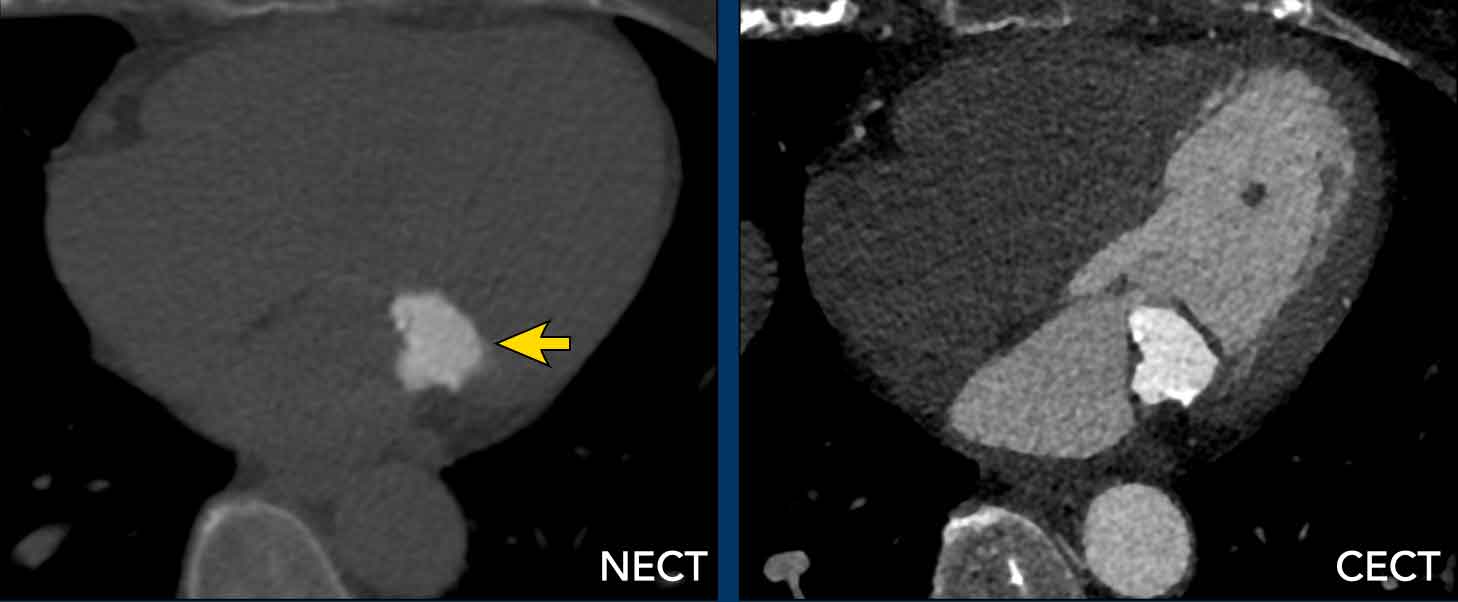
cMAC is a degenerative variant of mitral annular calcification, marked by liquefactive necrosis within a calcified annular mass.
It most commonly affects the posterior mitral annulus and is often an incidental finding in asymptomatic elderly patients.
Recognition is critical to avoid misdiagnosis as a cardiac tumor or abscess, which could lead to unnecessary investigations.
Rarely, it may serve as a source of embolic strokes or a nidus for infective endocarditis.
Example
Unenhanced CT and CTA in a patient with typical caseous mitral annular calcification.
cMAC (2)
Video of a patient with cMAC.
Cardiac tumors
Cardiac myxomas are the most common benign primary cardiac tumours in adults, although they remain rare overall.
75% arise in the left atrium, usually originating from the interatrial septum.
Many are detected incidentally on imaging, however, if symptomatic, they may present with obstructive cardiac symptoms, embolic events, and/or constitutional complaints related to cytokine release.
Their characteristic mobility and pedunculated attachment explains the possible intermittent obstruction of valvular inflow, particularly across the mitral valve.
Surgical resection is typically curative, although follow-up is recommended due to the possibility of recurrence.
Video example
A large hypoattenuating mass in the
right atrium.
Some mobility is seen on the 4-chamber cine series.
After
contrast administration no early enhancement.
Continue with the CT-video and late contrast MR-image...
CT-video and late phase MR
Heterogeneous late
enhancement is present.
Imaging findings are consistent with an atrial myxoma, which was proven after
surgical resection.
Myxoma (2)
Video example
Left atrial myxoma with intermittent transvalvular movement across the mitral valve.
Myxoma (3)
Images
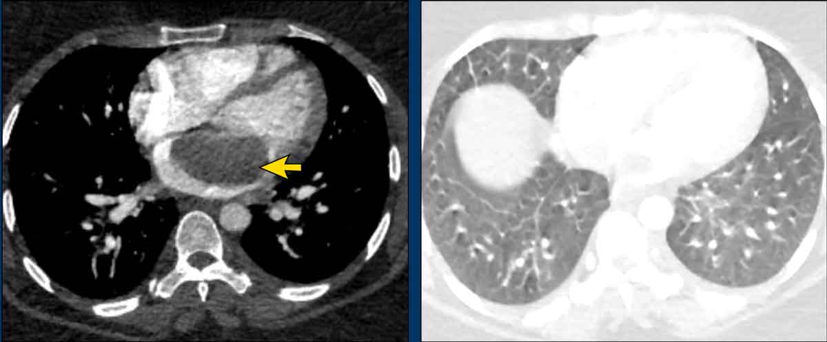
A patient presented to the ER with obstructive symptoms and was found to have an incidental left atrial myxoma.
Imaging revealed interstitial thickening due to cardiogenic edema and small pleural effusions (not shown).
Thrombus mimicking a myxoma
Video example
A right atrial mass was identified.
Although a myxoma was initially considered, MRI revealed no pedunculated connection to the septum but rather an attachment to an indwelling central venous catheter.
There was no early or late enhancement (not shown).
This supports the diagnosis of thrombus.
Lipoma
Cardiac lipomas are benign tumors composed of mature adipose tissue and can occur anywhere in the heart.
They are typically asymptomatic unless they grow large enough to cause obstruction, arrhythmia, or compression of adjacent structures.
On imaging, lipomas appear as well-circumscribed masses with macroscopic fat density and no internal enhancement.
These characteristic imaging features usually allow for a confident non-invasive diagnosis.
 Courtesy of Wouter Velthuis, UMC Utrecht
Rhabdomyoma
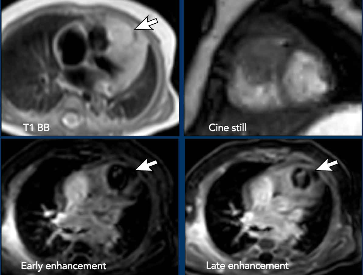
Cardiac rhabdomyomas are the most common benign primary cardiac tumors in children and are strongly associated with tuberous sclerosis complex (TSC). These mesenchymal hamartomatous lesions originate from striated muscle and most frequently involve the ventricular myocardium, often presenting as multiple intramyocardial nodules detected in utero or in young children.
Example
A cardiac mass in the right ventricle extended into the right ventricular outflow tract, appearing slightly hyperintense to the myocardium on T1-weighted images, with minimal heterogeneous enhancement on both early and late gadolinium-enhanced images. Histopathology confirmed a rhabdomyoma following a biopsy, which was performed to rule out other differential diagnoses such as fibroma, teratoma, and rhabdomyosarcoma.
Rhabdomyoma (2)
While most rhabdomyomas are clinically silent and demonstrate spontaneous regression over time, obstruction or arrhythmias may occur depending on size and location.
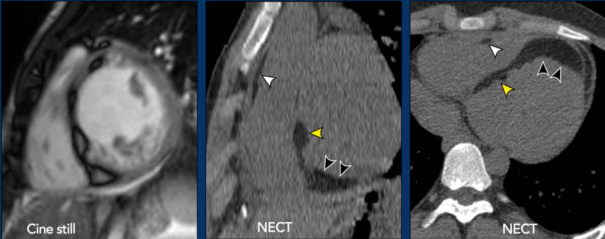
In older patients, fibrofatty replacement may be observed after involution.
Recognizing their typical pediatric presentation, multiplicity, spontaneous regression, and strong association with TSC is key to avoiding unnecessary interventions.
Images
Multifocal extensive
lipomatous replacement in
the myocardium, after regression of rhabdomyomas in a young adult TSC patient.
There is involvement of the interventricular septum (yellow arrowheads) extending into the
LV inferior wall (black arrowheads), and similar smaller lesions in the RV wall and RV
outflow tract (white arrowheads).
Fibroma
Cardiac fibromas are benign primary cardiac tumors that predominantly occur in infants and children but can also arise in adults.
Unlike rhabdomyomas, they do not regress spontaneously and may enlarge over time.
Typically located within the ventricular myocardium, they can cause ventricular arrhythmias, outflow obstruction, or heart failure.
Surgical resection is recommended for symptomatic patients, while asymptomatic lesions may be managed conservatively.
Example
Biopsy proven cardiac fibroma arising from the right ventricle myocardium, with homogeneous late gadolinium enhancement.
Papillary fibroelastomas (PFE)
Papillary fibroelastomas are the most common primary neoplasms of the cardiac valves, primarily affecting the aortic and mitral valves.
Their highly mobile, villous surface increases the risk of systemic embolization, with stroke being a recognized clinical presentation.
However, most cases are discovered incidentally.
Surgical excision is generally recommended due to their embolic potential.
Sarcoma
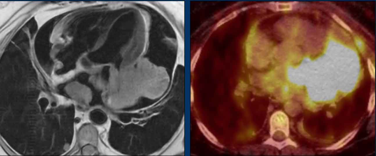
Cardiac sarcomas are the most common primary malignant heart tumors, though they remain extremely rare. Subtypes include angiosarcoma, liposarcoma, and undifferentiated pleomorphic sarcoma.
These tumors often involve multiple cardiac chambers, exhibit pericardial involvement, and invade adjacent structures, reflecting their aggressive and infiltrative nature. Patients typically present with non-specific symptoms such as obstruction or effusions, leading to delayed diagnosis. Rapid growth, local invasion, metastatic spread, and recurrence are common, resulting in a poor prognosis. Treatment often requires multimodal therapy, including surgical debulking, chemotherapy, and radiotherapy.
Video example
A large irregular,
lobulated mass with heterogeneous contrast enhancement, involving the left
atrium, the left ventricle wall and pericardium. There is a large pericardial effusion.
Continue with the video of the CT...
Sarcoma (2)
The video of the CT also shows the invasive character of this sarcoma.
Continue with the PET-CT image...
Primary cardiac lymphoma
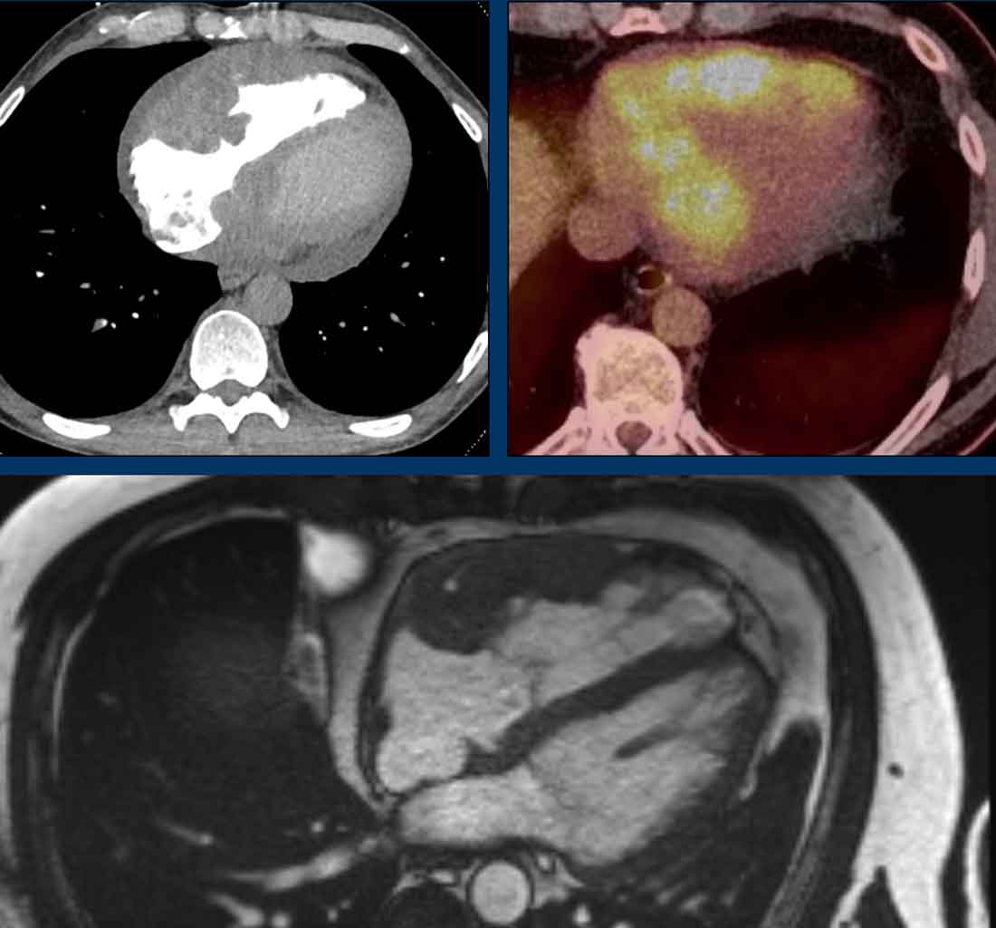
This is a rare form of extranodal non-Hodgkin lymphoma, most commonly of the diffuse large B-cell type.
It predominantly involves the right heart chambers, often extending along the pericardium as a soft-tissue mass.
Example
Multimodality imaging
findings in primary cardiac lymphoma, showing a diffuse mass at the right
atrium, AV-groove and ventricle with intense FDG uptake.
Despite an initial favourable response, relapse occurred
with eventual fatal disease progression.
Continue with the follow up images...
Primary cardiac lymphoma (2)
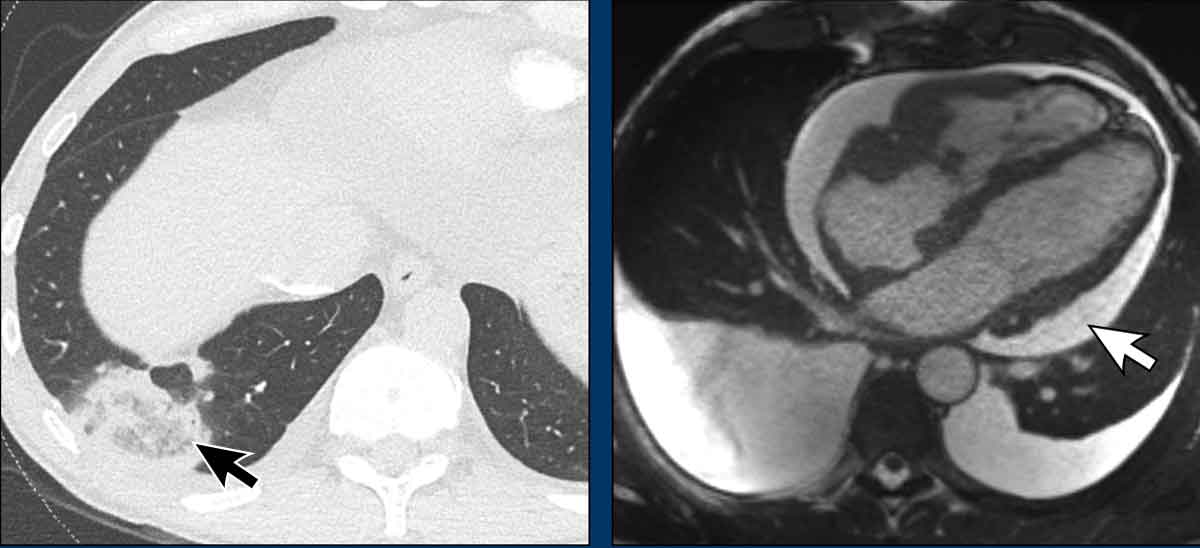
Images
Interval follow-up imaging shows pulmonary infarction due to emboli (black arrow).
There is significant pleural and pericardial effusion (white arrow).
The clinical presentation of primary cardiac lymphoma is typically non-specific, with symptoms such as arrhythmia, heart failure, pericardial effusion, or cardiac tamponade.
Unlike most primary cardiac tumors, it is potentially treatable with systemic chemotherapy, making early recognition critical.
Despite its aggressive imaging appearance, the prognosis can be favorable.
Metastases
Cardiac
metastases are by far the most common malignant tumors of the heart.
Clinical
presentation is variable and depends highly on lesion size and location.
Metastases may be incidentally seen on imaging in oncology follow-up, but are
more frequently detected at autopsy.
Cardiac metastatic
disease typically occurs in the setting of advanced-stage malignancy, most
commonly from lung and breast cancer, melanoma, and renal cell carcinoma, among
others.
Prognosis is generally poor, and treatment is usually palliative,
focusing on management of the underlying primary malignancy.
Example
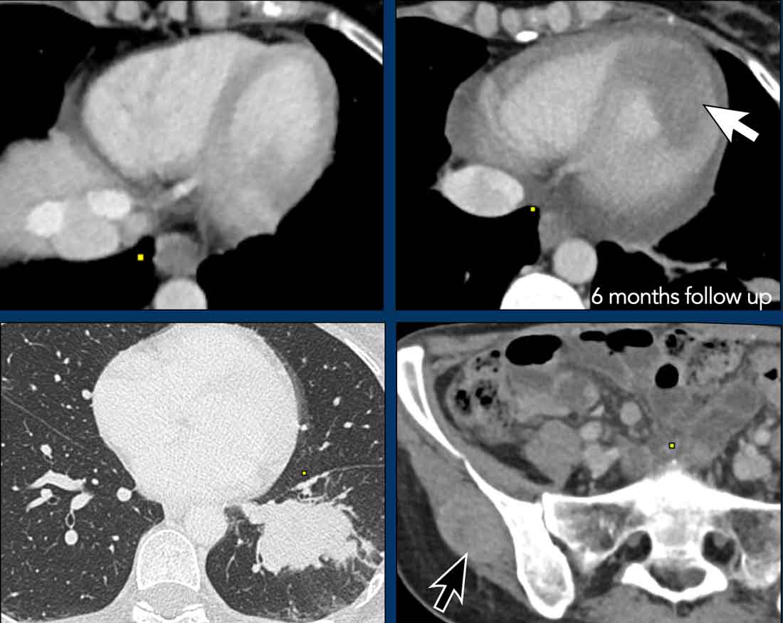
Cardiac metastatic
disease with a new large apical mass (white arrow) in oncologic follow-up for cervical
cancer.
There are also pulmonary, renal and soft tissue metastases (black arrow) present.
Metastases (2)
Images
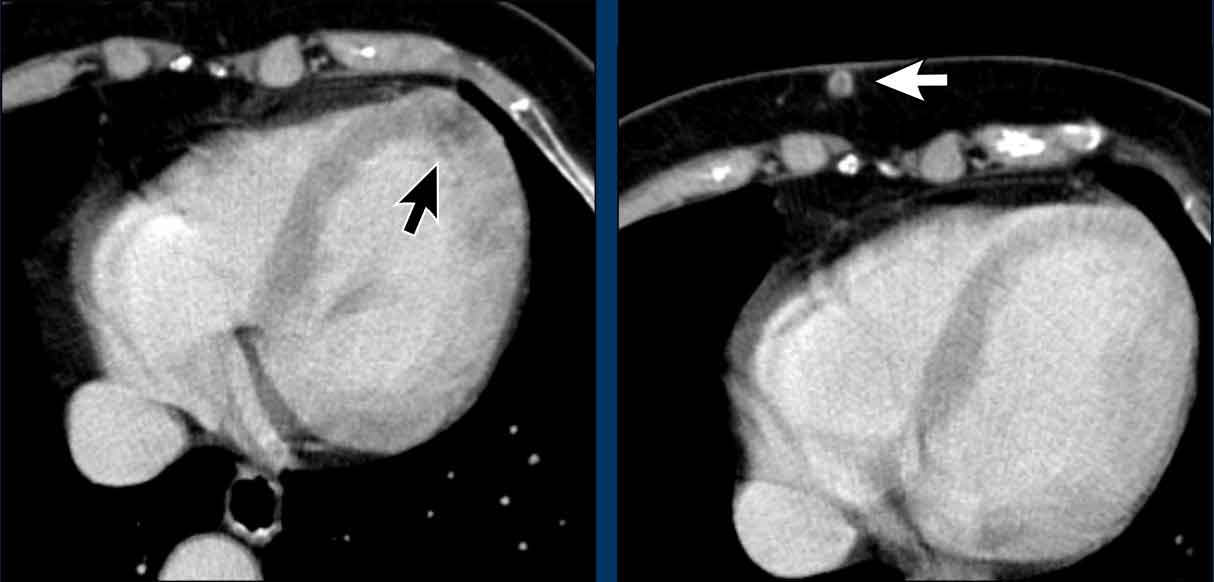
Subtle
left ventricular metastasis (black arrow) in a patient with renal cell cancer.
Note the synchronous
hypervascular soft tissue metastasis (white arrow) at a lower level.